
FIGURE 2.
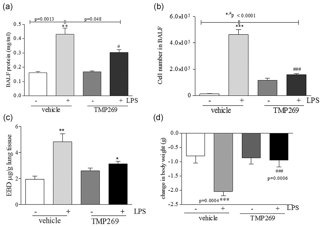
TMP269 pretreatment significantly attenuates LPS-mediated lung inflammatory processes in the murine model of ALI. (a) TMP269 (2.5 mg/kg), or saline (vehicle) was instilled intravenously 15 min before LPS (intratracheally) challenge. The protein content (mg/ml) was determined from the lavage fluid in the supernatants using bicinchoninic acid protein assay kit. Values are mean ± SEM, n = 4–6. **p < .005 vehicle versus LPS, #p < .05 LPS versus TMP269/LPS. (b) BALF was collected from all groups and it was centrifuged. Total numbers of cells were counted using a hemocytometer. Values are mean ± SEM, n = 4–6. ***p < .0001, **p < .001 vehicle versus LPS; *p < .05 LPS versus TMP269/LPS. (c) EBD-conjugated albumin was administered via tail vein 2 h prior the termination of the experiment. Then lungs were collected and EBD was measured. (d) TMP269 (2.5 mg/kg), or saline (vehicle) was instilled intravenously 15 min before LPS (intratracheally) challenge. Body weight was measured before and at the termination of the experiment. Mice lost weight after LPS exposure; values are mean ± SEM, n = 4–6. ***p = .0004 vehicle versus LPS; ###p = .0006 LPS versus TMP269/LPS. ALI, acute lung injury; EBD, Evans Blue Dye; LPS, lipopolysaccharide