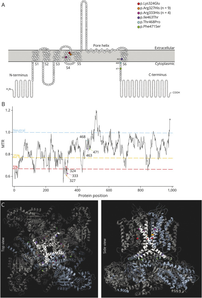
Figure 1. Location and Conservation of the 6 Unique De Novo KCNH5 Missense Variants.
(A) Schematic of Kv10.2 (EAG2), modified from Protter35 plot of Q8NCM2 (KCNH5_Human). Of the 6 transmembrane domains, S1-S4 make up the voltage-sensing domain, and S5-6 form the pore helix. The KCNH5 patient-specific variants identified in this study are indicated by their amino acid change. (B) Graph of missense tolerance ratio (MTR; y-axis) and protein position (x-axis), with variants indicated. MTR36 is a measure of protein-encoding cDNA sequence intolerance to missense variants. Variant positions with a value greater than 1 (blue line) is considered neutral; values below 1 are under constraint. Two variants (p.Arg327His and p.Arg333His) are in the 5th percentile of least tolerated missense alterations in the exome. The 5th and 25th percentiles are highlighted in red and yellow, respectively. (C) Locations of variants (color coded) are mapped onto the crystal structure of homotetrameric assembly of Kv10.1 (PDB5K7L); all variants are perfectly conserved between KCNH5 and KCNH1 and are mapped to the corresponding amino acid. The pore domain is highlighted in white, and a single tetrameric subunit is colored light blue. EAG = ether-a-go-go

