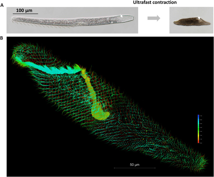
Fig. 1. The ultrafast contractile single-celled eukaryote, S. minus.
(A) Extended state (normal state, left) and contracted state (right) of S. minus. The white arrow indicates a groove on the surface of S. minus cell. (B) Three-dimensional image of the microtubule-based fibrillar bundles stained using tubulin–Atto 488 (73), imaged using super-resolution microscopy. Gradient colors were used for different Z-depth.

