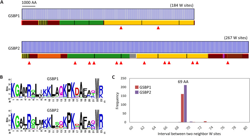
Fig. 3. Two giant proteins (GSBP1 and GSBP2) serve as spasmin-binding backbones.
(A) Features of GSBP1 and GSBP2 in S. minus. Blue vertical bar indicates the tryptophan (W) sites. Repeat regions are indicated by bars with different colors. The same color means repeat regions (separated by black vertical bars) with high sequence identity. Filled red triangles indicate coiled-coil regions. (B) Sequence logos show the motif of 23 amino acids (AA) around the tryptophan sites. (C) The 69–amino acid intervals between two neighboring tryptophan sites.

