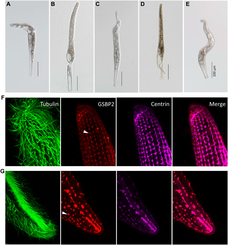
Fig. 6. RNAi of two giant proteins.
(A and B) Cells breaking down at anterior or posterior, respectively. (C and D) Cells showing distortion at anterior and posterior, respectively. (E) Distortion and twisting of entire cell. Scale bars, 100 μm. IF images showing the changes of mesh-like structure in GSBP2 RNAi cell using anti-GSBP2 antibody (rabbit). (F) Posterior region. (G) Anterior region.

