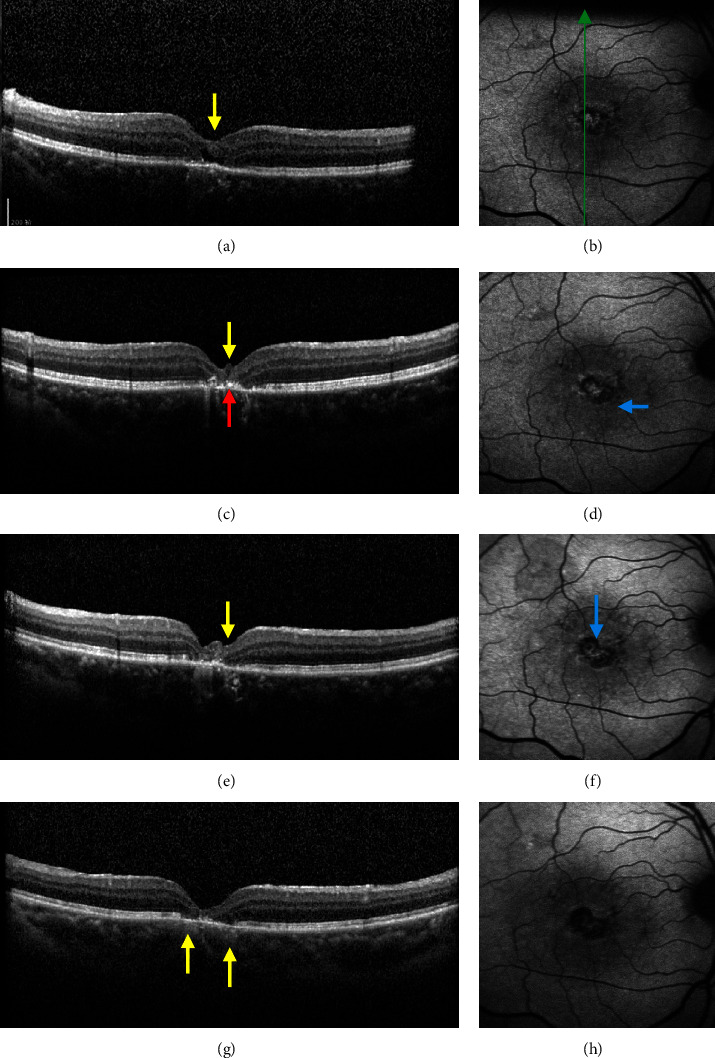
Figure 1.

Effect of inverted ILM flap technique on macular hole in IMH patients. Ocular SD-OCT and FAF images of one patient before surgery (a, b), one month after surgery (c, d), three months after surgery (e, f), and six months after surgery (g, h). The green arrow indicates the location and direction of OCT scanning; yellow arrows indicate protuberance with moderate signal intensity; red arrow indicates particles with high signal intensity; and blue arrows represent new spots with low signal intensity.
