
Figure 4.
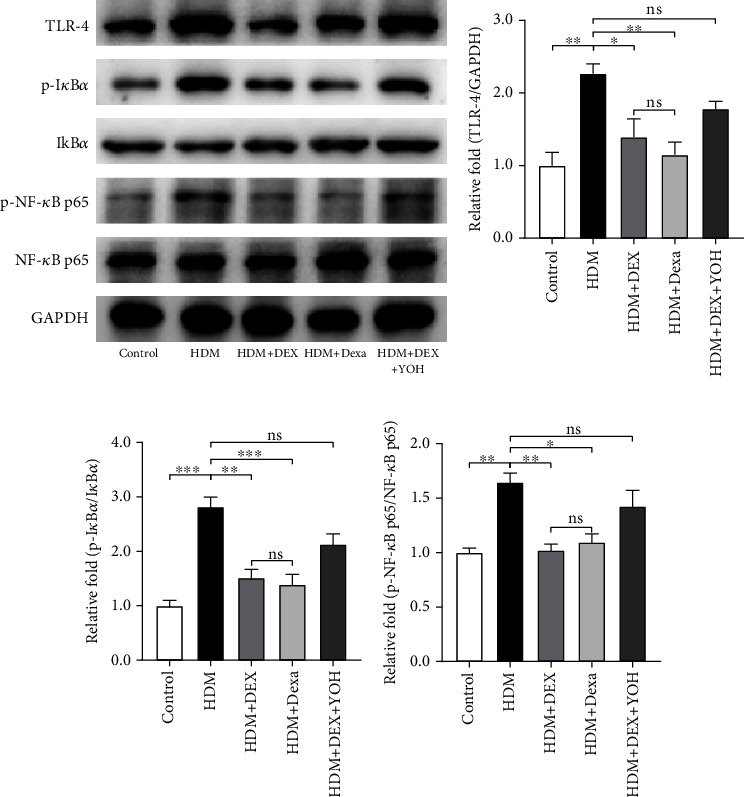
Effect of DEX on the expression levels of the TLR4/NF-κB signaling pathway in lung tissues. (a) Representative images of western blots of TLR4, phosphorylated- (p-) IκBα, IκBα, p-NF-κB p65, and NF-κB p65. (b–d) Densitometric analyses of TLR4 and the ratios of p-IκBα/IκBα and p-NF-κB p65/NF-κB p65 in lung tissues form each group. Data were presented as mean ± SEM (n = 3~5 animals). ∗P < 0.05, ∗∗P < 0.01, and ∗∗∗P < 0.001.