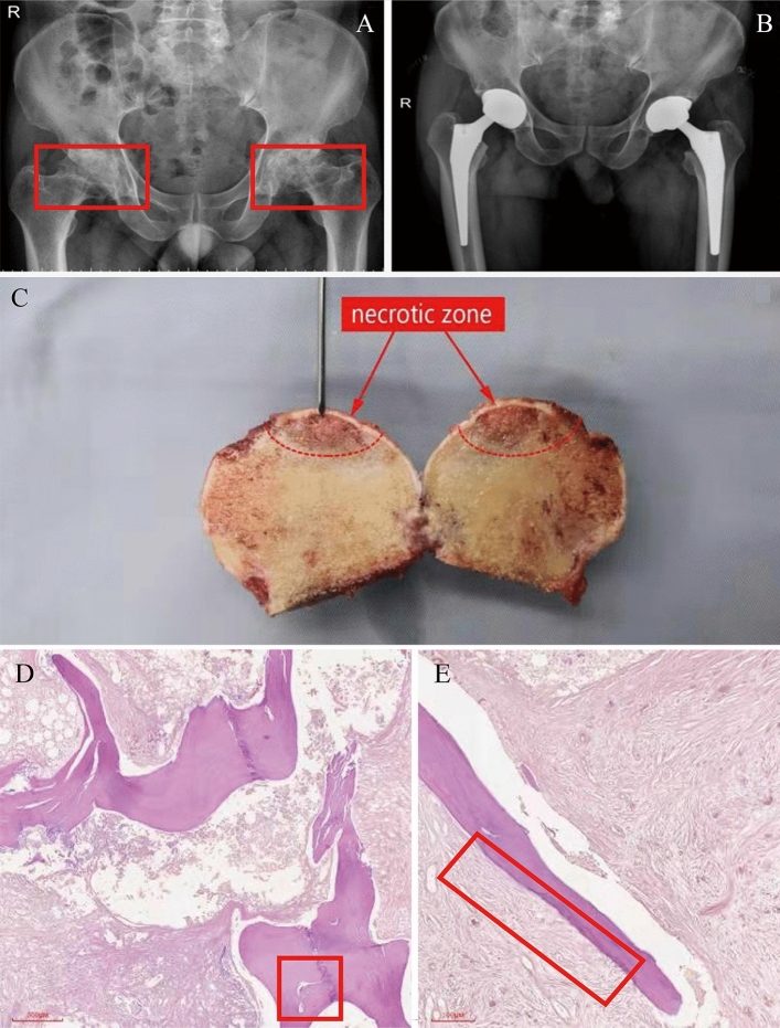
Figure 4.
Imaging system for clinical diagnosis. (A) Preoperative pelvic frontal radiograph (X-ray). Bilateral femoral heads were flat, with multiple cystic low-density shadows and narrowed joint space (red boxes on both sides) (B) Implanted prosthesis on pelvis orthographic film (X-ray) (C) Diseased tissue (with a red arrow pointing to necrosis). (D) Morphological abnormality in bone trabecular (left red frame) (40 ×). (E) Bone trabecular necrosis (right red box) (100 ×).

