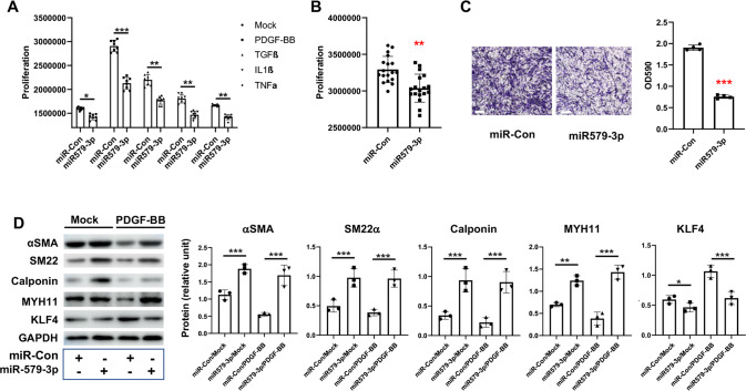
Fig. 2. miR579-3p suppresses AoSMC proliferation, migration, and de-differentiation.
A, B Proliferation assay. AoSMCs starved in basal medium (no FBS, 24 h) were transfected with miR-con or miR579-3p for 24 h. The medium was then changed to fresh basal medium with or without a cytokine (A) (50 ng/ml PDGF-BB, 20 ng/mL TGFβ1, 20 ng/ml TNFα, or 10 ng/ml IL1β) or full medium (B) for an additional 24 h before the CellTiter‐Glo viability assay. C Migration assay. AoSMCs were seeded in the Transwell insert, the lower chamber filled with full medium. Cells that migrated to the lower surface of the insert were imaged after a 24 h incubation. To quantify the migration, 33% (v/v) Acetic acid was added into the insert to elute the bound crystal violet, and then the eluent from the lower chamber was measured for absorbance (590 nm) using a 96-well microplate reader. Scale bar: 200 μm. D De-differentiation (Western blotting of SMC contractile proteins). AoSMCs were starved (no FBS, 24 h) and transfected with miR-con or miR579-3P for 24 h, and then treated with or without 50 ng/ml PDGF-BB for another 24 h. Quantification for A-D: Mean ± SD (A and B, see dot plots for the number of replicates) or mean ± SEM (C and D, n = 3 independent repeat experiments). Pairwise comparison was made through Student’s t-test. **P < 0.01, ***P < 0.001.

