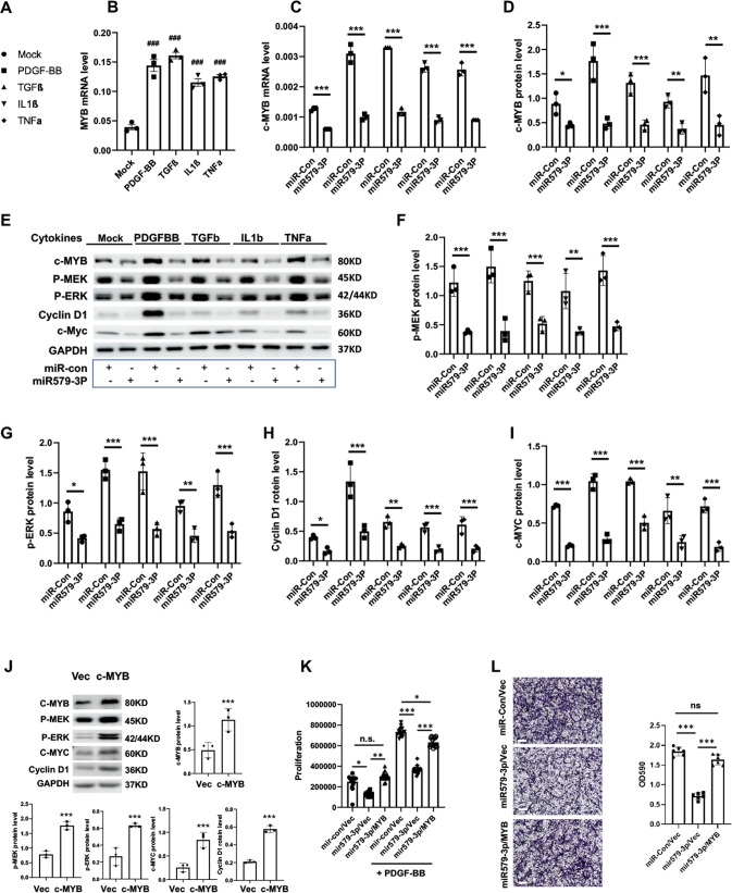
Fig. 3. miR579-3p represses the expression of c-MYB and proliferation/migration marker proteins.
AoSMCs starved in basal medium (no FBS, 24 h) were transfected with miR-con or miR579-3p for 24 h. The medium was then changed to fresh basal medium with or without a cytokine (50 ng/ml PDGF-BB, 20 ng/mL TGFβ1, 20 ng/ml TNFα, or 10 ng/ml IL1β) or full medium (migration assay) for an additional 24 h prior to various assays. A Lend of symbols (for B–I). B qRT-PCR showing upregulation of c-MYB mRNA by cytokines. C qRT-PCR showing negative regulation of c-MYB mRNA by miR579-3p. D–I Western blots showing that miR579-3P negatively regulates protein levels of c-MYB and proliferation/migration markers. Phospho-protein levels were normalized to the loading control GAPDH. J Western blots showing that c-MYB overexpression increases proliferation/migration markers. K Proliferation assay indicating that c-MYB overexpression rescues miR579-3P-mitigated AoSMC proliferation. PDGF-BB was included in the SMC culture. L Transwell assay indicating that c-MYB overexpression rescues miR579-3P-inhibited AoSMC migration. Scale bar: 200 μm. Quantification for A–L: Data are presented as mean ± SD (n = 3 replicates in B and C; n = 6 replicates in K and L) or mean ± SEM (western blots, n = 3 independent repeat experiments in D–J); ###P < 0.001 (compared to Mock control, the first bar), as analyzed with one‐way ANOVA followed by Bonferroni post hoc test. Pairwise comparison was made through Student’s t-test: *P < 0.05, **P < 0.01, ***P < 0.001.

