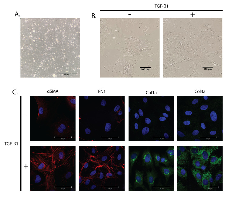
Figure 2. Cultured primary cardiac fibroblasts treated with TGF-β1.
(A) Bright-field image of attached neonatal rat cardiac fibroblasts on poly-L-lysine-coated dishes. Scale bar: 500 μm. (B) Bright-field image of primary cardiac fibroblasts with and without TGF-β1 treatment for 24 h. Scale bar: 100 μm. (C) Primary cardiac fibroblasts were treated with TGF-β1 for 24 h and stained with α-smooth muscle actin (α-SMA) as a marker for myofibroblasts, and fibronectin 1 (FN1), collagen 1a (Col1a), and Collagen 3a (Col3a) as fibrotic markers. Hoechst 33342 was used to stain the nuclei blue. Scale bar: 50 μm.

