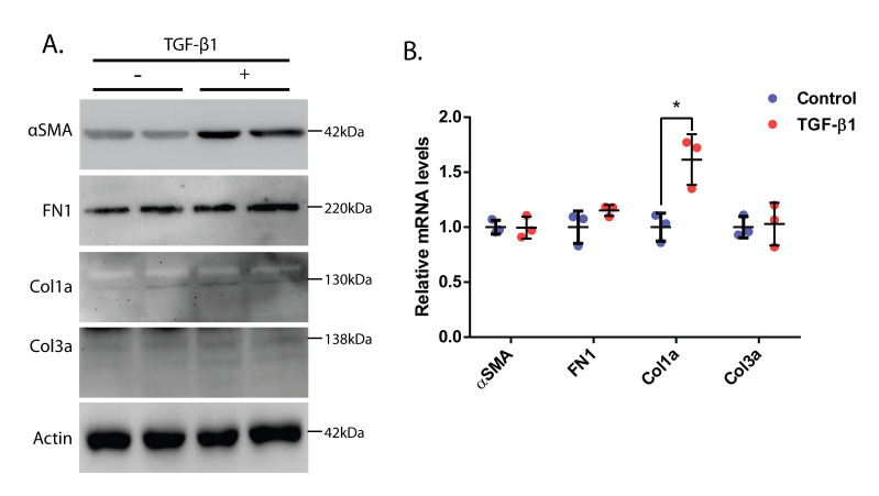
Figure 3. Molecular analysis of cardiac fibroblasts treated with TGF-β1.
(A) Western blot analysis of neonatal rat cardiac fibroblasts upon 24 h of TGF-β1 treatment. Α-SMA was used as a myofibroblast marker, and FN1, Col1a, and Col3a were used as fibrotic markers. Actin was used as the loading control. (B) Relative mRNA levels of α-SMA, FN1, Col1a, and Col3a in neonatal rat primary cardiac fibroblasts treated with TGF-β1 for 24 h. Actin has been used for normalization (as an internal control). Student’s t-test used for statistical analysis (*p ≤ 0.05). Data presented as mean ± S.D.

