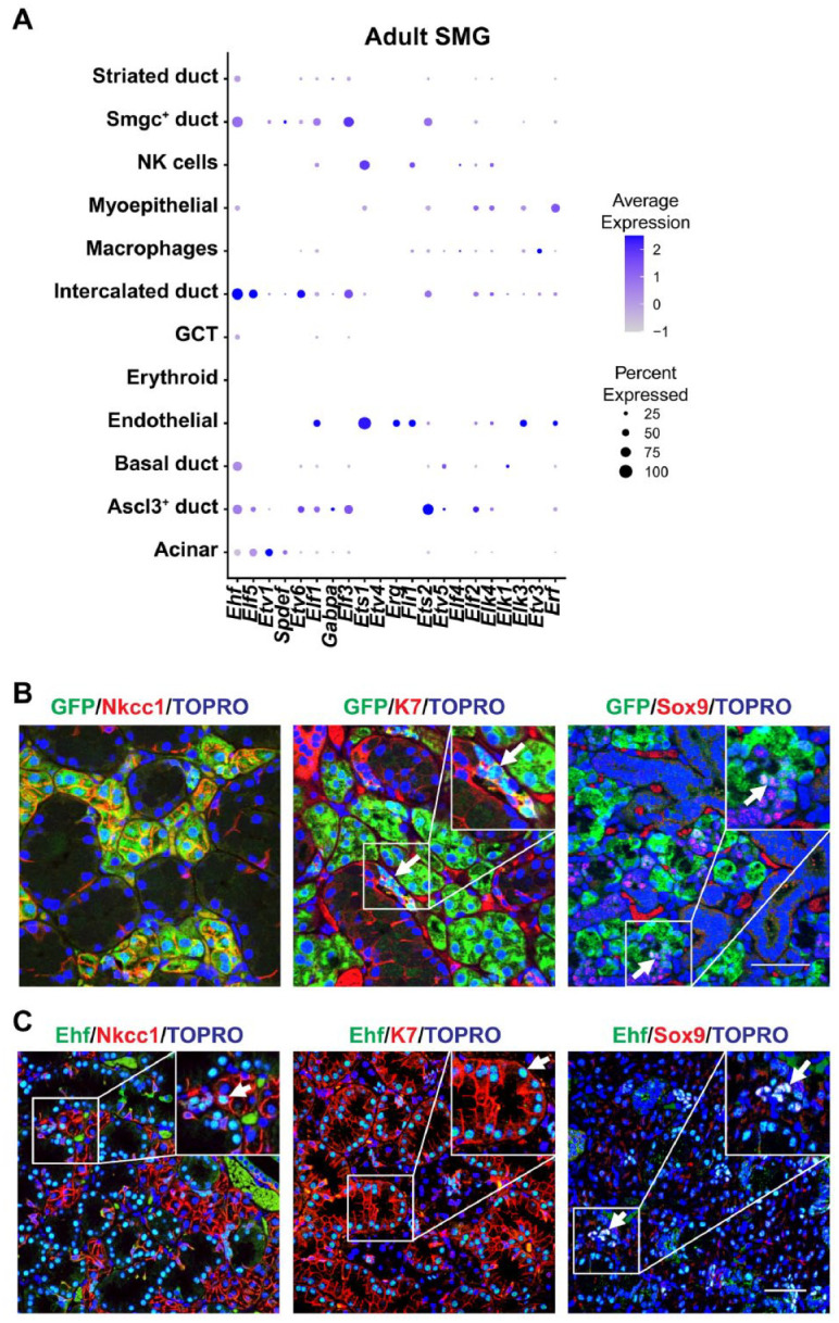
Figure 1.

Single-cell RNA sequencing analysis and protein expression profile of Elf5 and Ehf in mouse submandibular glands (SMGs). (A) Dot plot showing the scaled expression (dot color) and percent expression (dot size) of the ETS gene family members in each cell type in adult female SMG (GSE145268) (Min et al. 2020). (B) Immunofluorescence staining of Elf5–green fluorescent protein (GFP) (Pearton et al. 2011) transgenic mouse SMGs shows coexpression of GFP with acinar (Nkcc1), ductal (K7), and intercalated ducts (Sox9). Arrows highlight double-positive cells. (C) Ehf protein expression profile in 10-wk-old adult male mouse SMG showing colocalization of Ehf with the broad ductal (K7) and specific intercalated ductal marker (Sox9). Arrows indicate Ehf colocalization with indicated cell markers. Yellow: green and red colocalization; pink: red and blue (nuclei) colocalization; turquoise: green and blue colocalization; white: green, red, and blue colocalization. Scale bar: 50 µm. GCT, granular convoluted tubule.
