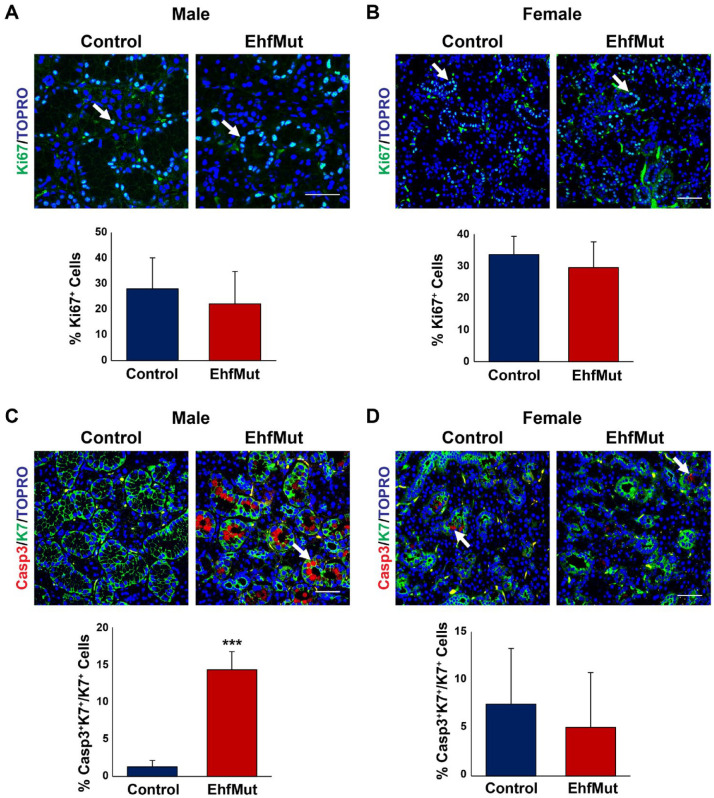
Figure 4.
Ductal cells undergo apoptosis in EhfMut mice. (A) Expression and quantification analysis of cell proliferation based on Ki67 expression (white arrows) show no differences between control and male EhfMut glands and (B) female control and mutant glands. (C) Expression and quantification analysis of cleaved caspase-3 (Casp3) reveals increased apoptosis in the K7+ ducts of the male EhfMut submandibular gland (SMG) compared to control mice. Arrows indicate the K7+Casp3+ double-positive cells. (D) Expression and quantification analysis of cleaved Casp3 reveals similar levels of apoptosis in the ducts of the female EhfMut SMG compared to control mice. Arrows indicate ductal cells undergoing apoptosis. Data are represented as mean ± standard deviation (SD) (n = 5). ***P < 0.001. Scale bar: 50 µm.

