
Figure 2. Cardiac development.
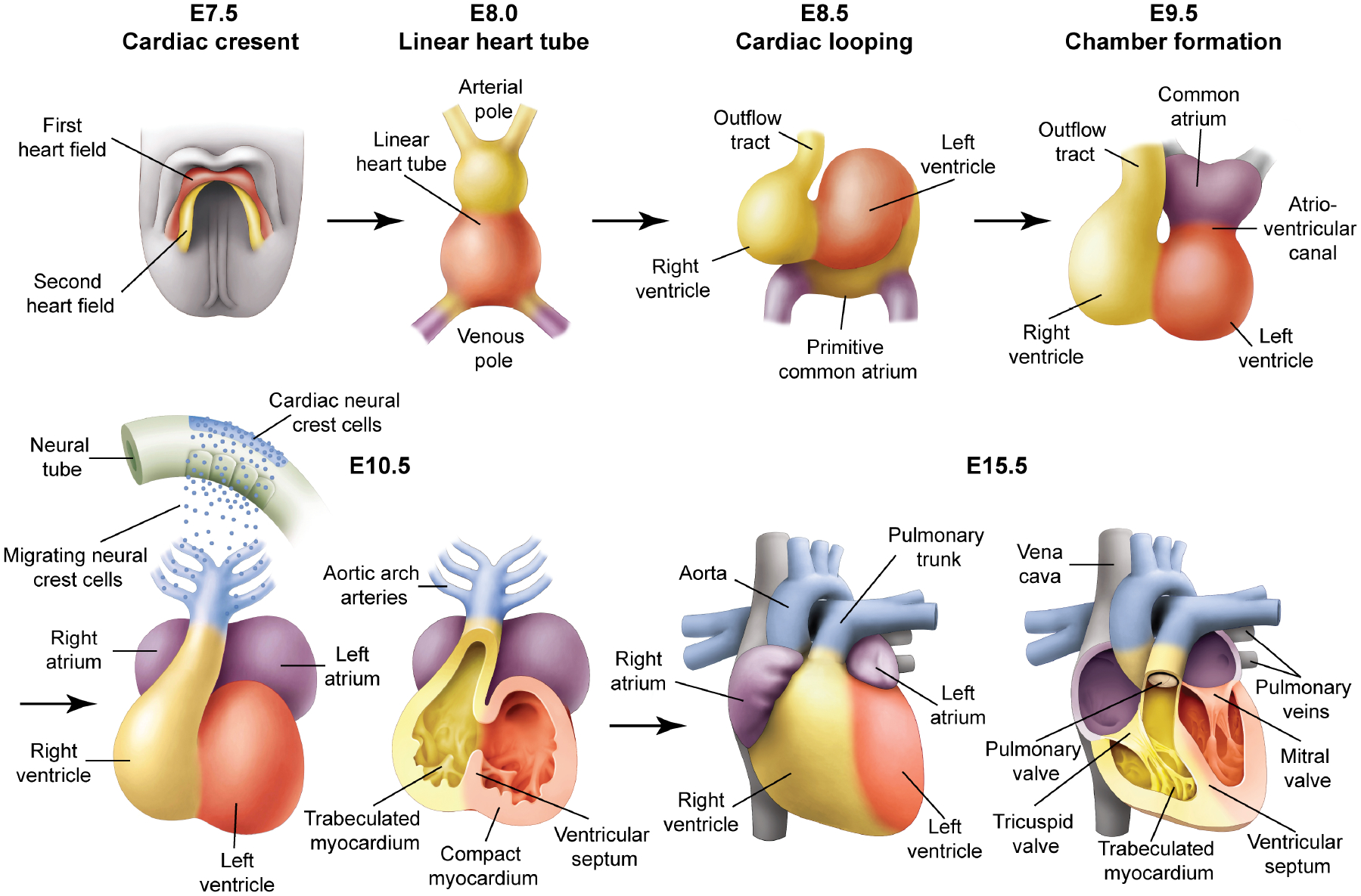
The heart originates from mesodermal cells in the primitive streak. During gastrulation, cardiac progenitors migrate to the splanchnic mesoderm to form the cardiac crescent. At E7.5 in the mouse, the cardiac crescent can be divided into two heart field lineages based on differential gene expression and their respective contribution to heart, a first heart field (red) and a second heart field (yellow), which is located posteriorly and medially to the first heart field. At E8.0, the linear heart tube is present. At E8.5, the looping is associated with uneven growth of cardiac chambers. The outflow tract is at the arterial pole and the inflow tract and primitive atria are at the venous pole. By E9.5, the common atrium has moved superior to the ventricles and is separated by a distinct atrio-ventricular canal. By E10.5, cardiac neural crest cells from the dorsal neural tube migrate via the pharyngeal arches to the cardiac outflow tract. Further cardiac development involves a series of septation events and myocardial trabeculation that result in a mature four-chambered heart integrated with the circulatory system depicted at E15.5.