
Figure 3. Schematic overview of mammalian vascular development.
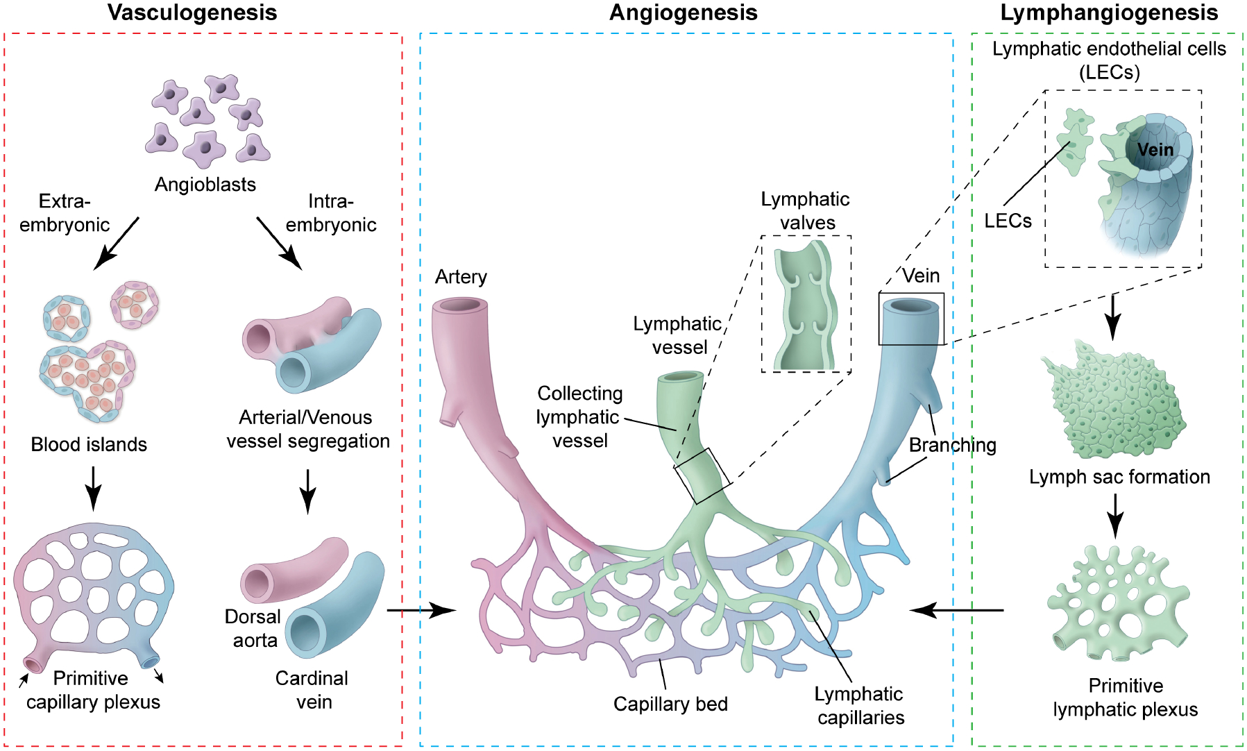
Vascular endothelial progenitor cells (angioblasts) are derived from mesodermal cells during gastrulation. These mesodermal cells have potential to give rise to both blood and endothelium. However, their fate is restricted once they have emigrated into extra-embryonic tissue (extra embryonic ectoderm, yolk sac and allantois) and intra-embryonic tissues (embryonic ectoderm). In extra-embryonic tissue, these angioblasts aggregate to form blood islands. Fusion of blood islands leads to the formation of a honeycomb-shaped primitive capillary plexus. However, in intra-embryonic tissue, angioblasts aggregate and directly form the dorsal aorta and cardinal vein, without a vascular plexus intermediate. The primitive vascular plexus along with dorsal aorta and cardinal vein undergo remodeling to form a mature circulatory system. Lymphatic endothelial cells (LECs) are specified in embryonic cardinal veins at defined locations. Under the influence of Vegfc, produced by the mesenchymal cells surrounding the cardinal veins, LEC precursors lose their polarity, delaminate from the veins, migrate and aggregate to form the primary lymph sacs. Furthermore, sprouting, remodeling and expansion of the lymph sacs and primitive lymphatic plexus lead to the formation of mature peripheral lymphatic vasculature. There are two types of lymphatic vessels: collecting lymphatic vessels and lymphatic capillaries. Lymphatic capillaries are thin-walled, blind-ended vessels that absorb interstitial fluid and transport it to the collecting lymphatic vessels. Collecting lymphatic vessels are surrounded by smooth muscle cells and have bicuspid valve to facilitate unidirectional lymphatic flow.