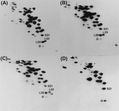
FIG. 1.
Association of SRA (protein D) with the 30S ribosomal subunit. Electropherograms from RFHR 2-D PAGE analysis of the ribosomal proteins in total cell extracts (A), crude ribosomes (B), high-salt-washed ribosomes (C), and 30S subunits (D) prepared from W3110 cells grown to the stationary phase. Protein D (SRA), S21, L32, and L33 spots are indicated.

