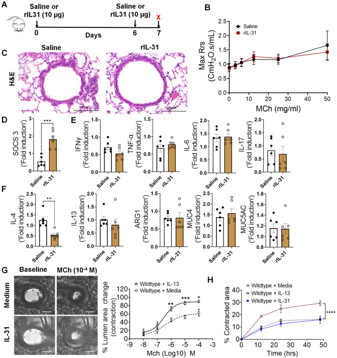
Figure 6. IL-31 is dispensable for the induction of AHR, inflammation, and Th2 responses.
(A) Schemata showing intratracheal administration of IL-31 or saline in wild-type mice. (B) Measurement of resistance with increasing doses of methacholine (MCh) in wild-type mice treated with saline or IL-31 using FlexiVent. Data are shown as mean ± SEM, n = 6/group. The data is representative of two independent experiments with no statistical significance between groups. (C) Representative images of hematoxylin and eosin -stained lung sections from wild-type mice treated with IL-31 or saline. Images were captured at 20x magnification, scale bar 100 μm. (D) Quantification of IL-31-induced SOCS3 gene expression in the whole lung tissue of wild-type mice treated with saline or IL-31. Unpaired two-tailed Student’s t-test was used. ***P < 0.001. (E and F) Quantification of inflammation-associated gene transcripts IFN-γ, TNFα, IL-6, and IL-17 and Th2-associated gene transcripts including IL-4, IL-13, ARG1, MUC4, and MUC5AC in whole lungs of wild-type mice treated with saline or IL-31. Unpaired two-tailed Student’s t-test was used and no significance found between groups, n=6/group. (G) Representative images of precision cut lung sections (PCLS) from wild-type mice treated with or without IL-31 (500 ng/ml) for 24 h. Airway contractility was measured in response to MCh (10−4 M) compared to baseline diameter. The percent of airway lumen area contraction with increasing doses of MCh was calculated for saline and IL-31-treated PCLS from wild-type mice. Two-way ANOVA test, n=5–8/group. (H) The percent contraction of collagen gels embedded with airway smooth muscle cells from wild-type mice that were treated with media, IL-13 (50 ng/ml) or IL-31 (500 ng/ml). The percent contraction was measured at different time points compared to baseline. Two-way ANOVA was used, n=4/group.

