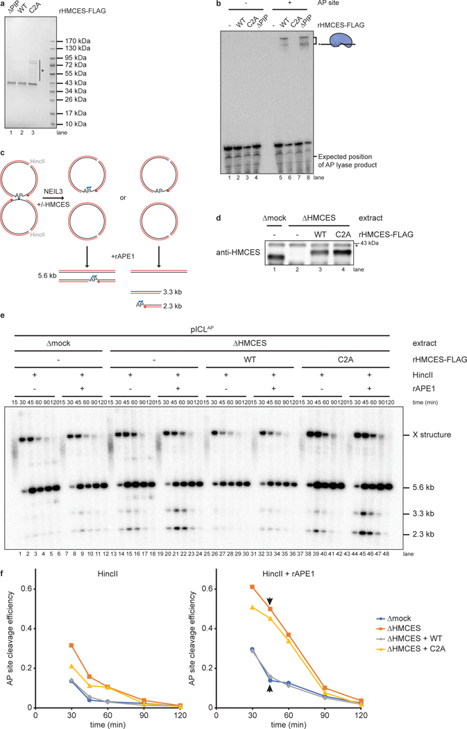
Extended Data Fig. 1. An HMCES-DPC shields AP sites.
a, Purified recombinant FLAG-tagged Xenopus laevis HMCES proteins were resolved by SDS-PAGE and visualized by staining with InstantBlue. rHMCESΔPIP harbors W321A and L322A mutations that disrupt a conserved PIP-box that was previously show to mediate interaction with PCNA3. Asterisk, contaminating bands.
b, A 5’ end radiolabeled 20mer oligonucleotide with a single deoxyuracil (-AP site) or AP site (+AP site) was incubated with rHMCES proteins shown in (a) for 60 min. Samples were then resolved on a denaturing polyacrylamide gel and visualized by autoradiography. Reactions contained 1 nM oligonucleotide and 50 nM rHMCES.
c, Schematic of species produced by digestion of pICLAP replication intermediates with HincII and APE1. Digestion with HincII generates a 5.6 kb linear plasmid species while additional AP site cleavage by APE1 is expected to generate 2.3 kb and 3.3 kb species.
d, HMCES immunodepletion. The extracts used in the reactions shown in e were blotted for HMCES. Asterisk, non-specific band.
e, pICLAP was replicated with [α−32P]dATP in the indicated egg extracts (shown in d). Samples were treated with proteinase K, phenol:chloroform extracted, and digested with HincII or with HincII and APE1. Digested DNAs were resolved on a native agarose gel and visualized by autoradiography. X structures indicate HincII-digested plasmids before ICL unhooking.
f, Quantification of APE1 cleavage efficiency for the reactions shown in e. Cleavage efficiency was quantified as the intensity (Int) of 2.3 kb and 3.3 kb fragment bands in each lane divided by the total intensity of linear species bands ([Int2.3kB + Int3.3kB]/[Int2.3kB + Int3.3kB + Int5.6kB]). The efficiency of HMCES-DPC formation in mock-depleted extract was estimated by subtracting the extent of rAPE1 cleavage in mock-depleted extract from the extent of cleavage in HMCES-depleted extract at 45 min (arrowheads), when the absolute signal resulting from rAPE1 cleavage is maximal.

