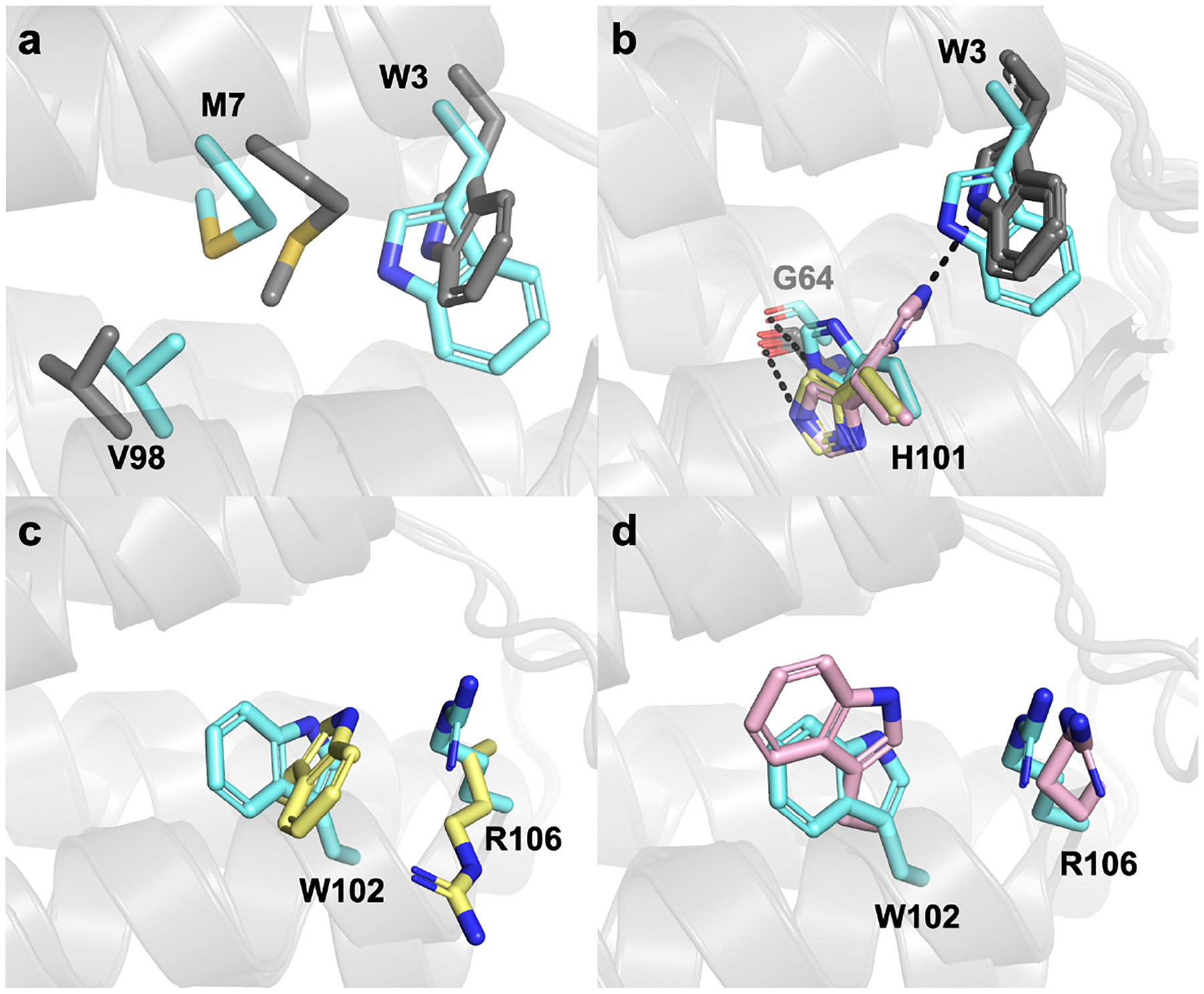
Figure 5.

Structural alignments of the design model (cyan) to the ApoCyt crystal structure. (a) Alignment of the design model to Monomer A (grey), focusing on residues that are structurally the same across different ApoCyt monomers. (b) Alignment of the design model to Monomers A and C (Conformation 1, yellow) and Monomers B and D (Conformation 2, pink) highlighting the alternate conformations of H101. (c) Comparison of residues 102 and 106 in the design model to Conformation 1. (d) Comparison of residues 102 and 106 in the design model to Conformation 2.
