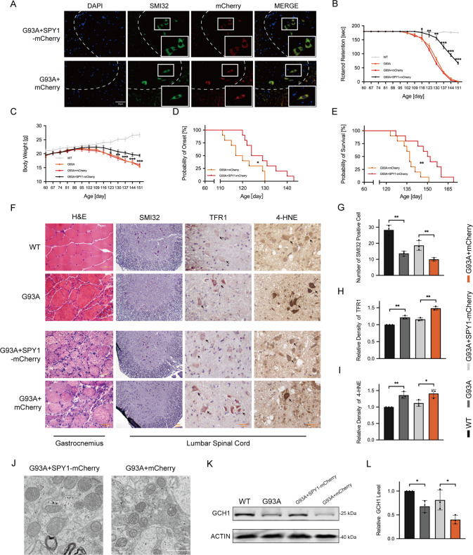
Fig. 6. SPY1 plays a protective role in hSOD1G93A mice by resisting neuronal ferroptosis.
hSOD1G93A transgenic mice at 60 d were injected with AAV9-hSyn-SPY1-mCherry and AAV9-hSyn-mCherry. A The overexpressed SPY1 in green MNs of lumbar spinal cord stained with SMI32 was verified by autofluorescence of mCherry at 90 d. Scale bar, 50 μM. B, C The decreased body weight and rotarod test were recorded (n = 10). D, E Kaplan–Meier survival analysis for probability of onset and survival (n = 10). F Images of immunohistochemical staining in end-stage mice. First column for the gastrocnemius muscle stained with H&E. The residue for the lumbar spinal cord tissue immunohistochemically stained with SMI32, TFR1, and 4-HNE. Scale bar, 50 μM. G–I Quantification for the number of SMI32 positive cells and relative density of TFR1 and 4-HNE (n = 3). J Electron microscopy for mitochondrial changes of the lumbar spinal cord in hSOD1G93A mice with overexpression of SPY1 and control. Scale bar, 1 μM. K, L Western blots and quantification for the effect of mutant SOD1 and overexpressed SPY1 on the expression of GCH1 in lumbar spinal cord (n = 3). Values represent mean ± SD. Statistical analysis by one-way or two-way ANOVA followed by Tukey’s post hoc tests. *p < 0.05, **p < 0.01, ***p < 0.001.

