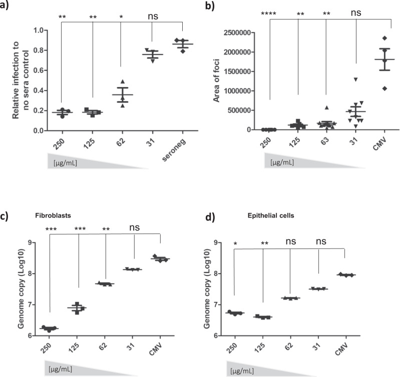
Fig. 6. Anti-AD-6 antibodies limit cell-associated spread of CMV.
a–c To measure spread of cell-associated virus in HFFs, cells were infected with Merlin-IE2-GFP (MOI = 0.01). At 1 dpi and 5 dpi, anti-AD-6 antibody was added at the indicated concentrations (plus control normal rabbit sera in A. At 10 dpi, cells were then analysed by IF or DNA-qPCR for viral spread. Percentage of infected cells (a) was measured by anti-IE stain counterstained with nuclei stain and counted by automated fluorescence microscopy (n = 3) and expressed relative to infection seen in cells incubated with no sera. Alternatively, the area of each individual plaque identified from randomly chosen images from the three independent experiments analysed in a was measured using Fiji software (b). Alternatively, total DNA was harvested, and CMV genome copies per 106 cells assessed by qPCR (c; n = 3 independent experiments reporting on biological replicates). d ARPE-19 cells were infected with pentamer positive BAC derived Merlin-IE2-GFP (MOI = 0.01), and total DNA harvested 20 dpi and analysed for viral genome copy number per 106 cells n = 3; independent experiments reporting on biological replicates. For all analyses (a–d) P values were calculated by a Kruskal–Wallis test with Dunn’s multiple-comparison test where appropriate. ****P < 0.0001; ***P < 0.001; **P < 0.01 *P < 0.05; ns P > 0.05. In all cases (a–d), the error bars represent 1 standard deviation from the mean.

