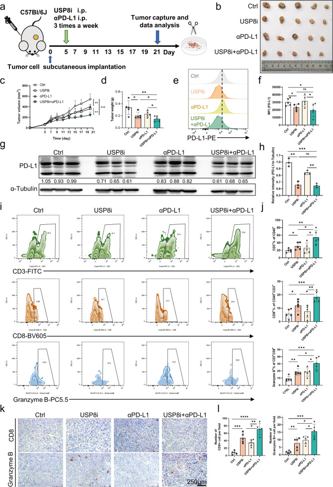
Fig. 5. Pancreatic tumor growth in vivo is suppressed effectively using a USP8 inhibitor combined with αPD-L1.
a Protocol schematic of the combination of USP8 inhibitor and αPD-L1 therapy for mice implanted subcutaneously with KPC cells (5 × 10⁵). b Photographs of tumors removed from mice treated with the USP8 inhibitor, αPD-L1, or their combination (n = 5). c Curves showing the tumor growth in mice treated with the USP8 inhibitor (100 μg/mouse), αPD-L1 (200 μg/mouse), or their combination (n = 5). d The statistical plot of tumor weights of the four groups (n = 5). e, g PD-L1expression assessed using flow cytometry and western blotting in a subcutaneous tumor model, statistical results (f, h) shown. i, j Tumor-infiltrating lymphocytes (TILs) assessed using flow cytometry and the statistical analysis of the results (n = 5). k, l Representative images of IHC staining and quantification of TILs (n = 5). Scale bars = 250 μm. The results are shown as means ± SD from representative experiments in (c, d, f, h, j and l). The data represent three independent experiments. *p < 0.05, **p < 0.01, ***p < 0.001, ****p < 0.0001 assessed via a two-tailed t test; ns: not significant.

