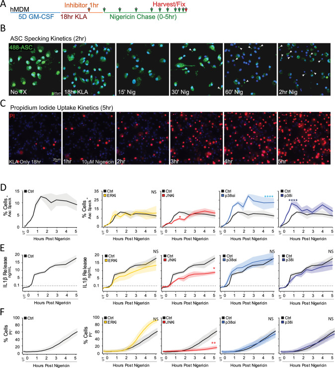
Fig. 1. JNK Inhibition Blunts Inflammatory Cell-Death and IL-1β Secretion in hMDM.
A hMDM were subjected to an 18 h 100 nM KLA extended prime regimen prior to inflammasome triggering with 10 µM nigericin. B, C Inflammasome formation was evaluated by endogenous ASC speck imaging (B; White arrows) while pyroptotic cell permeation was evaluated by propidium iodide (PI) uptake (C; Red stain) to analyze kinetic differences between the two events. Blue indicates Hoechst-stained nuclei. D–F Following 18 h 100 nM KLA priming, individual MAPK inhibitors (ERK [10 µM U0126], JNK [30 µM JNK-IN-8], p38α [10 µM SB203580], and pan-p38 [30 µM doramapimod]) were applied for 1 h before inflammasome triggering with 10 µM nigericin to examine roles for MAPKs in ASC specking (D), IL-1β release (E), and cell permeation (F). D–F Shading represents standard deviation from the mean. Two-way ANOVA followed by Tukey’s multiple comparison test; *p < 0.05, **p < 0.01. Comparable results were obtained in parallel studies with U937 cells (see Figs. S1 and S2). B–F Data shown are representative of at least three independent experiments.

