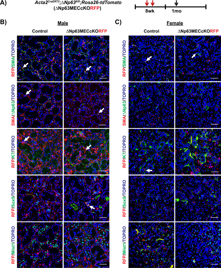
Fig. 4. Lineage tracing of MECs 1 month post ΔNp63 deletion in this cell population under homeostatic conditions.
A Experimental timeline used for lineage tracing of myoepithelial cells upon deletion of ΔNp63. Animals were administered TAM to simultaneously induce ΔNp63 specific deletion in the MECs and irreversibly label MECs with RFP expression. Animals were traced for 1 month and SMGs were analyzed. Immunostaining of the B male and C female control and ΔNp63MECcKORFP glands. Arrows indicate co-localization of RFP-positive cells with various epithelial cell markers. Myoepithelial: SMA, ΔNp63, Ductal: K7, Sox9, Acinar: Mist1. Scale bar: 50 µm.

