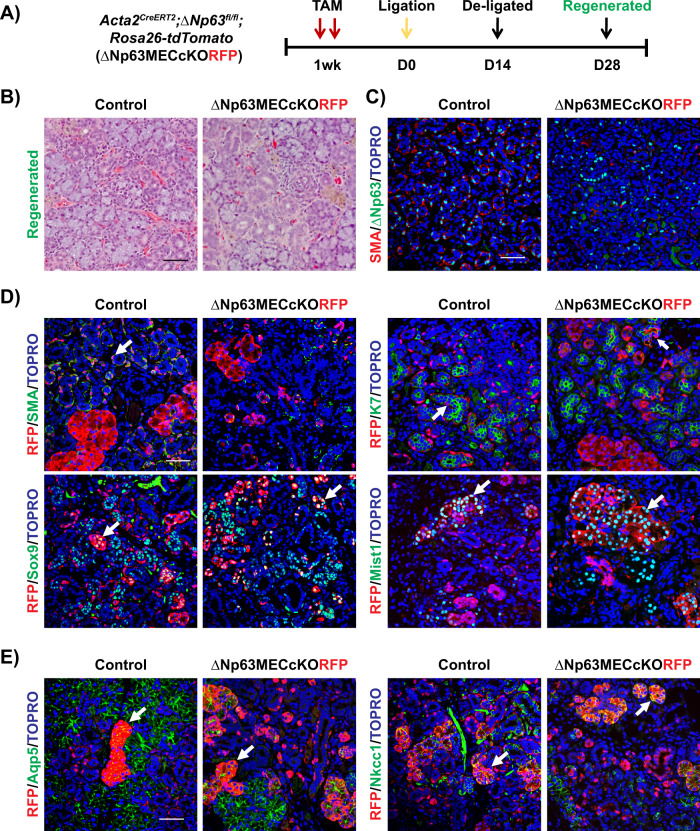
Fig. 6. Contribution of ΔNp63 ablated MECs to SMG regeneration following severe injury.
A Experimental timeline used to collect the regenerated SMGs of the control and ΔNp63MECcKORFP mice. B Histological analysis of the regenerated female control and ΔNp63MECcKORFP glands. C Immunostaining images of regenerated KO glands stained with the MEC markers SMA and ΔNp63. D Immunochemical analysis shows that RFP co-localizes with K7, Sox9 (ductal marker), and Mist1 (acinar marker) in both the control and ΔNp63MECcKORFP regenerated SMGs. RFP+ cells represent the progeny of the SMA+ myoepithelial cells. E RFP+ cells co-localize with Aqp5 and Nkcc1 (acinar markers) in the control and ΔNp63MECcKORFP-regenerated SMGs. White arrows indicate co-localization of RFP and specific cell population markers. Scale bar: 50 µm.

