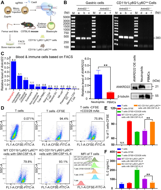
Figure 3.
Knockout of Ankrd22 enhanced the immunosuppressive activity of PMN-MDSCs. (A) Diagram of the construction of the Ankrd22-knockout C57BL/6 mouse model. (B). Bone marrow was collected from KO mice and WT mice to prepare single-cell suspensions, and then CD11b+Ly6G+Ly6Clow cells were sorted by FACS. PCR and agarose gel electrophoresis were used to detect Ankrd22. M: marker. n=3. Ankrd22 in gastric epithelial cells was used as a positive control. (C). The HPA database showed the expression level of ANKRD22 among human blood and immune cells based on flow sorted. Human peripheral blood neutrophils and mononuclear cells were extracted following the instruction of human peripheral blood neutrophil/mononuclear cell isolation kit, and the expression of ANKRD22 was measured by RT-qPCR and western blotting. The lysate of the ANKRD22-overexpressing (OE) 293 T cells as a positive control. n=3, **p<0.01. Two-sample Student’s t-test. (D, E) FCM was used to detect the percentage of proliferating CFSE-labeled T cells after coculture with CD11b+Ly6G+Ly6Clow cells which were treated with 100 ng/mL GM-CSF and 100 ng/mL IL-6 for 96 hours. CFSE fluorescence intensity of T cells were showed. n=3, **p<0.01. Two-sample Student’s t-test. (F). The content of IL-2 in the supernatant of cocultured cells was detected by ELISA. n=3, **p<0.01. Two-sample Student’s t-test. CFSE, carboxyfluorescein diacetate succinimidyl ester; FACS, fluorescenceactivated cell sorting; FCM, flow cytometry; HPA, Human Protein Atlas; PMN-MDSCs, polymorphonuclear myeloid-derived suppressor cells; WT, wild type.

