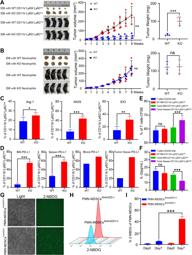
Figure 4.
Knockout (KO) of Ankrd22 promoted the proliferation of ovarian cancer cells by enhancing the immunosuppressive ability of polymorphonuclear myeloid-derived suppressor cells (PMN-MDSCs) in transplanted tumor mouse models. (A) 5×10⁶ ID8 cells/mouse mixed with 10×106 BM-derived CD11b+Ly6G+Ly6Clow cells from Ankrd22+/+ or Ankrd22-/- C57BL/6 mice, were suspended in Matrigel and injected into the axilla of WT C57BL/6 mice. Tumor diameters were measured with a Vernier caliper once a week for 8 weeks. The growth of subcutaneous tumors in mice was recorded. At the end of the experiment, the subcutaneous tumor weight was determined. n=4, **p<0.01, ***p<0.001. Two-sample Student’s t-test. (B). 5×10⁶ ID8 cells/ mouse were mixed with 10×106 neutrophils from mouse peripheral blood and injected into the axilla of WT C57BL/6 mice. Tumor diameters were measured with a Vernier caliper once a week for 8 weeks. The growth of subcutaneous tumors in mice was recorded. At the end of the experiment, the subcutaneous tumor weight was determined. n=4, NS: p>0.05. Two-sample Student’s t-test. (C). Subcutaneous tumor tissues were collected from KO and WT mice, and intracellular Arg-1, iNOS and IDO on CD11b+ Ly6G+Ly6Clow cells were detected by flow cytometry (FCM) after stimulation with 81.0 nM PMA and 1.34 µM ionomycin. n=4, *p<0.05, **p<0.01, ***p<0.001. (D) FCM was used to detect PD-L1 on CD11b+Ly6G+Ly6Clow cells derived from the bone marrow, spleen, peripheral blood or subcutaneous tumor tissues of the tumor-bearing mice in WT and KO groups. ***p<0.001. Two-sample Student’s t-test. (E.) FCM was used to detect the percentage of proliferating carboxyfluorescein diacetate succinimidyl este (CFSE)-labeled T cells after coculture with CD11b+Ly6G+Ly6Clow cells from mice tumor tissues without treatment of 100 ng/mL GM-CSF and 100 ng/mL IL-6 for 96 hours. CFSE fluorescence intensity of T cells were showed. n=3, **p<0.01. Two-sample Student’s t-test. (F). The content of IL-2 in the supernatant of cocultured cells was detected by ELISA. n=3, **p<0.01. Two-sample Student’s t-test. (G–I). CD11b+Ly6G+ Ly6Clow cells from the tumor-bearing mice were cultured with DMEM containing 2-NBDG under 100 ng/mL GM-CSF and 100 ng/mL IL-6 stimulation for 7 days. The intensity of green fluorescence in the PMN-MDSCs was detected by immunofluorescence microscopy. The FCM results show the percentage of PMN-MDSCs with FITC fluorescence. n=3, ***p<0.001. BM, bone marrow; FITC, fluorescein isothiocyanate.

