Figure 6.
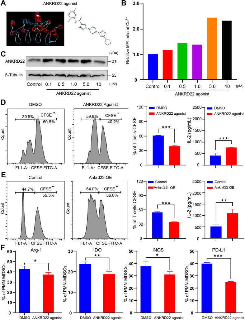
An ANKRD22 agonist reversed the immunosuppressive activity of polymorphonuclear myeloid-derived suppressor cells (PMN-MDSCs). (A) Schematic diagram of ANKRD22-activating small-molecule compounds. (B) Human THP-1 cells were cocultured with agonist at concentrations of 0, 0.1, 0.5, 1, 5 and 10 µM for 24 hours. The mean fluorescence intensity of Ca2+ was measured to evaluate whether the small-molecule compound was effective. (C) Colorectal cancer LS-174T cells were cocultured with agonists at concentrations of 0, 0.1, 0.5, 1, 5 and 10 µM for 24 hours. The expression level of ANKRD22 was detected by western blotting. (D) BM-derived CD11b+Ly6G+Ly6Clow cells from wild-type (WT) mice, stimulated with 100 ng/mL GM-CSF and 100 ng/mL IL-6 (PMN-MDSCsAnkrd22+/+), were cocultured with actived carboxyfluorescein diacetate succinimidyl ester (CFSE)-labeled spleen-derived T cells in the presence of 1.0 µM ANKRD22-activating small-molecule compound for 96 hours. DMSO was added to the FCM culture medium as a control. Flow cytometry (FCM) showed the effect of PMN-MDSCs on T-cell proliferation after addition of the activating small-molecule compound. ELISA was used to detect IL-2 concentration (pg/mL) in the supernatant of PMN-MDSCs cocultured with T cells. n=3, ***p<0.001. Two-sample Student’s t-test. (E) PMN-MDSCsAnkrd22+/+ were infected with recombinant lentivirus overexpressing Ankrd22. FCM shows the effect of PMN-MDSCs on T-cell proliferation after infection. ELISA was used to detect the IL-2 concentration in the cell supernatant of PMN-MDSCs cocultured with T cells. n=3, **p<0.01, ***p<0.001. Two-sample Student’s t-test. (F) PMN-MDSCs were treated with an ANKRD22 agonist for 72 hours in vitro, and the results showed that the levels of Arg-1, iNOS, IDO and PD-L1 were reduced in PMN-MDSCs which were with ANKRD22 agonist. n=3, *p<0.05, **p<0.01, ***p<0.001. Two-sample Student’s t-test. BM, bone marrow.

