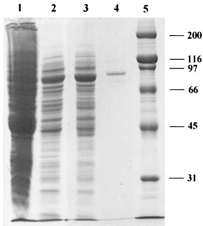
FIG. 2.
SDS-PAGE of phosphoketolase preparations from B. lactis. Lanes: 1, crude extract; 2, the sample after DEAE-chromatography; 3, the sample after the Mono Q column; 4, the enzyme after the Superdex 200 column; 5, protein standard. Numbers indicate the molecular mass (in kilodaltons) of the standard proteins. The gel (10% polyacrylamide) was Coomassie stained.

