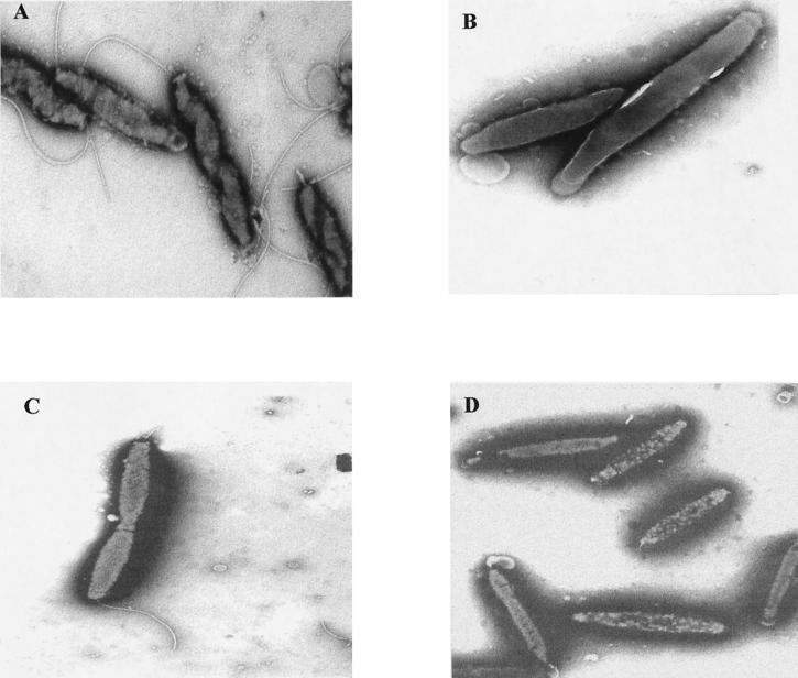
FIG. 3.
Transmission electron microscopy of the wild-type C. jejuni strain NCTC 11168 (A) compared to the rpoN (B), fliA (C), and flgR (D) mutants. Transmission electron microscopy was performed by scraping cells from plates grown overnight on MH agar at 37°C for 24 h under microaerophilic conditions. Cells were suspended in 50 μl of 1.5% (wt/vol) sodium phosphotungstate (pH 7.0), and a small drop of the suspension was applied to Formvar-coated copper grids. Excess suspension was removed with the edge of a filter paper, and negatively stained cells were visualized in a JEOL 1200EX 80-kV transmission electron microscope.

