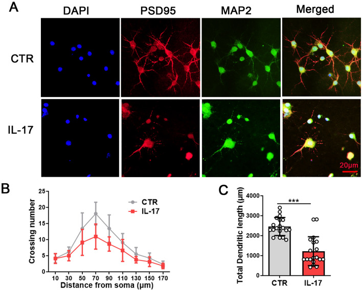
Fig. 2.
IL-17 induces neuronal toxicity in primary hippocampal neurons. Mouse primary hippocampal neurons were treated with DMSO for the control group and IL-17 for the IL-17 group for 9 d. Changes in neuronal morphology were measured by immunofluorescence staining with anti-PSD95 (red), anti-MAP2 (green) antibodies, and co-labelled with DAPI (blue). Representative images (20×magnification) after treatment (A), Sholl analysis (B), quantitative analyses of dendritic length (C), N = 20 hippocampal neurons. Data are presented as the mean ± SD. p value significance is calculated from a T-test. ***p < 0.001 vs the control group. For western blotting original images, please see Additional file 1, and Adobe Photoshop software (2021) was used to cut the parts of interest from the original images

