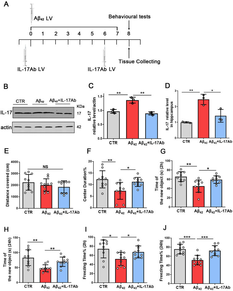
Fig. 3.
Inhibition of IL-17 alleviates Aβ42-induced cognitive deficits. Flow chart for the experiments (A). IL-17 levels were detected by western blotting using specific antibodies, and actin was used as a loading control (B). Intensity analysis of IL-17 levels (C). N = 3. ELISA to measure the levels of IL-17 (D), N = 3. The open-field test showed the total distance covered (E) and the time of center duration (F) of the three groups. NORT showed the time spent exploring new objects at 2 h (G) and 24 h (H). The contextual fear conditioning test determined the freezing time at 2 h (I) and 24 h (J). N = 10 for independent experiments. Data are presented as the mean ± SD. p value significance is calculated from a one-way ANOVA test, *p < 0.05, **p < 0.01 and ***p < 0.001 vs the Aβ42 group. For western blotting original images, please see Additional file 1, and Adobe Photoshop software (2021) was used to cut the parts of interest from the original images

