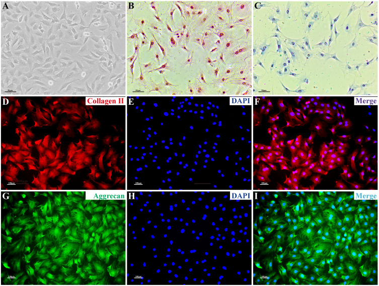
Figure 3.
NPC phenotype. (A) Morphological observation of NPCs. (B) NPCs stained with H&E; the nuclei are blue and the cytoplasm is pink. (C) After toluidine blue staining, the acidic aggrecan in the cytoplasm was visualized by blue staining, with the darker color observed closer to the nucleus. (D–I) Immunofluorescence staining: collagen II and aggrecan showed red and green fluorescence, respectively, with stronger fluorescence intensity observed closer to the nucleus; the nucleus showed blue fluorescence. Scale bars: 50 μm in (A–C), 100 μm in (D–I).
Abbreviation: NPC, nucleus pulposus cell.

