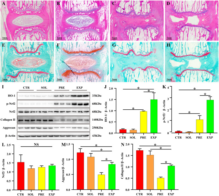
Figure 8.
ADR effectively inhibits static mechanical pressure-induced IDD. H&E staining of sagittal sections of the IVD showed that the disks in the CTR group (A) and SOL group (B) had good morphology, with a normal intervertebral space height. Additionally, the fibrous rings were pinkish in color and neatly arranged in an annular stratification without fibrous ring fracture or disorder. The central part of the nucleus pulposus was blue-purple and oval in shape, full in volume, and rich in extracellular matrix, and its internal type II collagen and aggregated proteoglycans were filled in lattice-like intracellular chambers. The nucleus was stained blue and the cell pulp was stained pink. The thickness of the upper and lower cartilage endplate was higher, and transparent chambers of different sizes were visible within the cartilage endplate and filled with abundant collagen and cartilage endplate cells. The disc height was significantly lower in the PRE group (C) and the EXP group (D) compared to the control and solvent groups, and the cartilage endplate was significantly thinner. The volume of the hyaline compartment within the cartilage endplate was significantly reduced, and the collagen content was significantly decreased, with inflammatory cell infiltration observed within it. In the PRE group, the fibrous rings were disorganized, with fissures and fractures inside the rings, and the nucleus pulposus was heavily resorbed, leaving only a small portion of the volume, with no internal cellular structure and a transparent appearance. In the EXP group, the volume of the nucleus pulposus tissue was significantly larger than that in the pressure group, although the fibrous rings were also disorganized, fissured, and fractured. The sagittal sections of intervertebral discs stained with safranine O-fast green FCF cartilage (E–H), (E) CTR group, (F) SOL group, (G) PRE group, (H) EXP group) showed similar results to those of H&E staining. (I–N) The expression of HO-1 and p-Nrf2 was significantly increased (P < 0.05) and the expression of collagen II and aggrecan was significantly decreased (P < 0.05) in the PRE and EXP groups compared to those in the CTR and SOL groups. HO-1, p-Nrf2, collagen II, and aggrecan expression were significantly increased in the EXP group compared to the PRE group (P < 0.05). Values measured are presented as the mean ± standard deviation, n=3 for each group. *P < 0.05.
Abbreviations: ADR, andrographolide; IDD, intervertebral disc degeneration; IVD, intervertebral disc; CTR, control; PRE, pressure; SOL, solvent; EXP, experimental; HO-1, heme oxygenase-1; Nrf2, NF-E2-related factor 2.

