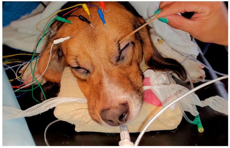
Figure 1.
The image shows the position of the color-coded needle EEG leads used in this study. The electrode R1 (white color) was positioned at the Fp2 in the human 10–20 EEG system. The R2 lead (green color) was positioned between F4 and F8 location. The L1 (blue color) lead was positioned at the Fp1 location, and the L2 lead (red color) was positioned between F3 and F7 location. The ground (CB, yellow color) and the reference (CT, black color) electrodes were placed on the mid-sagittal line in the central and the cranial position, respectively. The image also shows the use of a cotton swab for assessing the dog’s eye reflexes (palpebral and globe position).

