
Figure 3.
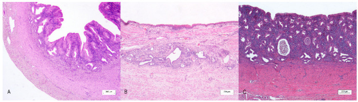
Uterus masculinus, histological findings: (A) Case number 3: The wall of the uterus masculinus with a three-layer appearance. Bar 200 µm. (B) Case number 2: Longitudinal section of the mucosal wall with squamous epithelium. Bar 200 µm. (C) Case number 1: Moderate lymphoplasmacytic and neutrophilic inflammation of the mucosal layer of the canine uterus masculinus. (A–C): Under 4× magnification, bar 200 µm, hematoxylin–eosin stain.