
Figure 4.
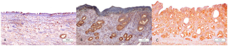
Uterus masculinus, immunohistochemical findings: (A) Case number 3: Multifocal-cytoplasmic AMH staining in the simple columnar epithelium of the uterine mucosa. Bar 100 µm. (B) Case number 2: Diffuse-negative immunolabelling of the uterine squamous epithelium and simultaneous strong glandular expression of AMH. Bar 100 µm. (C) Case number 1: Diffuse-strong glandular and epithelial AMH immunolabelling. (A–C): Under 10× magnification, bar 100 µm.