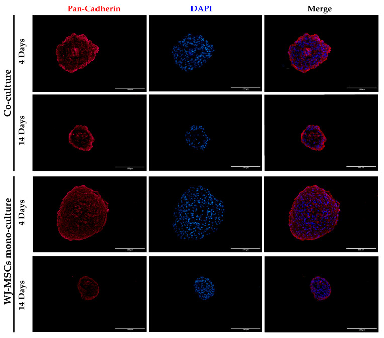
Figure 6.
Representative images of WJ-MSC mono-culture and co-culture spheroids at early (4 days) and late (14 days) time points stained with Pan-Cadherin (red) and DAPI for nuclei staining (blue). Cells were observed using a Nikon Inverted Microscope (Nikon Instruments, Tokyo, Japan), and images were acquired with a Digital Sight camera DS-03 using the imaging software NIS-Elements 4.1 (Nikon Corporation, Tokyo, Japan). Scale bar = 100 µm. Magnification = 20×.

