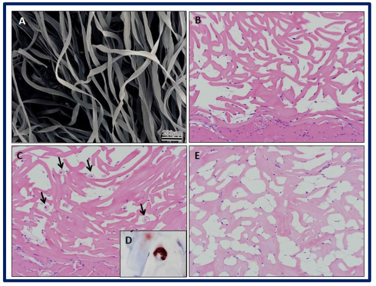
Figure 24.
SEM micrograph showing the (A) FSA scaffolds. Histological examination of implant specimens of FSA scaffolds at different weeks (Hematoxylin and Eosin staining). (B) Week 1: Note the presence of stained spindle cells attached to scaffold fibers. (C) Week 2: Black narrows indicate brown-like adipocytes within the scaffold, with multiple cytoplasmic lipid droplets. Note the presence of some small spherical cells close to scaffold fibers. No inflammatory reaction is observed. (D) Week 2: Multiple cytoplasmic lipid droplets within the cells are stained with Oil Red O staining. (E) Week 4: Multiple white-like adipocytes within scaffold bulk are observed, with large cytoplasmic lipid droplets. Bibliography consulted [196].

