
Figure 1.
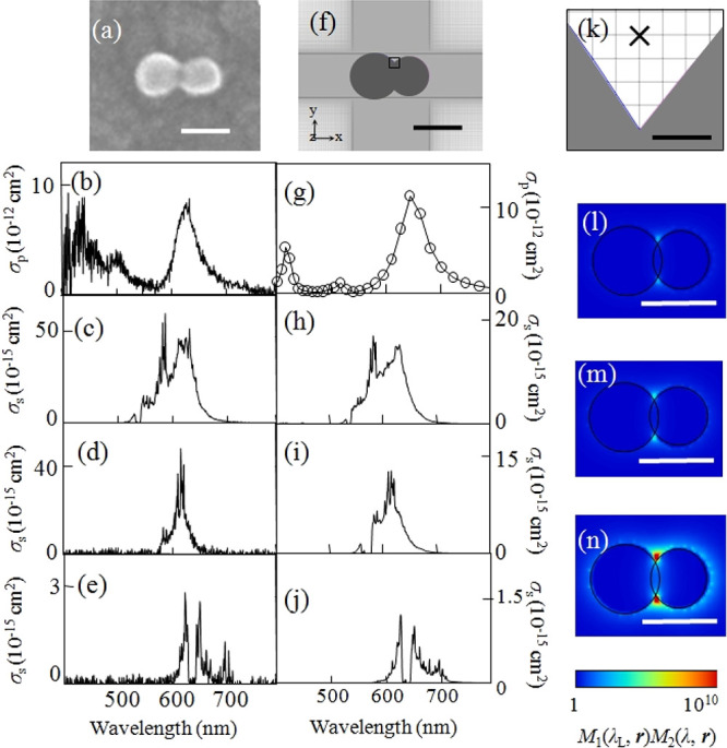
(a) SEM image of a silver NP dimer. (b) Experimental plasmon resonance spectrum and experimental resonant SERS spectra excited at (c) 532, (d) 561, and (e) 633 nm. (f) Modeled structure of a silver NP dimer in the FDTD calculation. (g) Calculated plasmon resonance spectrum and calculated SERRS spectra excited at (h) 532, (i) 561, and (j) 633 nm. (k) Magnified view in the vicinity of the crevice of the model structure in (f). Spatial distribution of the calculated SERRS-EM EF excited at (l) 532, (m) 561, and (n) 633 nm. Circularly polarized incident light (532, 561, and 633 nm), mesh size (0.2 nm), and refractive index (1.3) of the surrounding medium were selected for the FDTD calculation. The scale bars in panels a, f, and l–n are 50 nm, while that in panel k is 0.5 nm. Reproduced with permission from ref (51). Copyright 2010 American Physical Society.