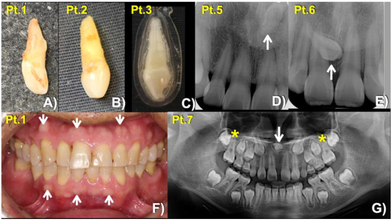
Figure 2.
Mesiodens phenotypes. Extracted mesiodens of (A) patient 1, (B) patient 2. (C) patient 3. (D,E) Periapical radiographs show (D) Patient 5—Inverted mesiodens (arrow). (E) Patient 6—Unerupted mesiodens (arrow). (F) Patient 1—Buccal exostoses (arrows). (G) Panoramic radiograph showing inverted mesiodens (arrow) and unseparated roots of the maxillary first permanent molars (asterisks).

