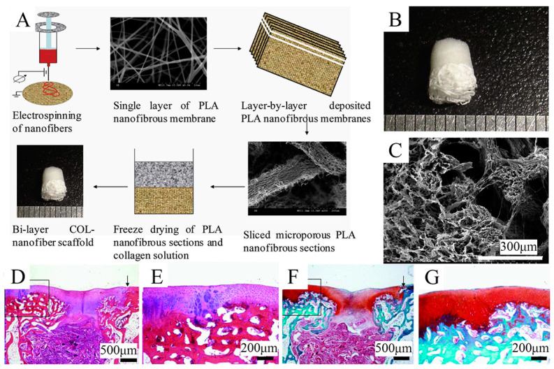
Figure 4.
Schematic representation of the combination of freeze-drying and 3D printing techniques used to fabricate the gradient scaffold. Reproduced with permission from Ref. [134]. Copyright 2013, Elsevier. (A) Fabrication process of COL-nanofiber scaffolds. (B) Macroscopic images of the COL-nanofiber scaffold showing obvious differences between two layers. (C) SEM images of the interface between two layers in the bi-layer scaffolds. (D,E) H&E staining of samples at 6 weeks after surgery. (F,G) Safranine O staining of samples at 6 weeks after surgery.

