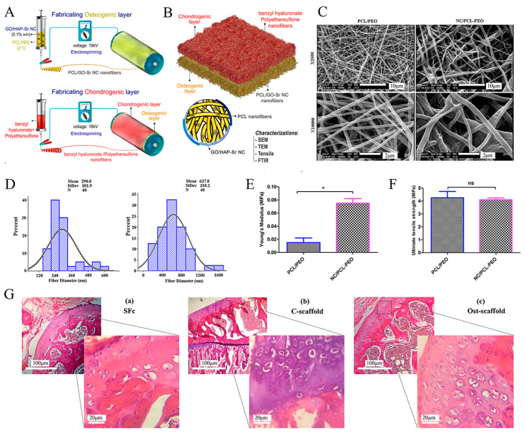
Figure 5.
Schematic illustration of a bilayer composite nanofibrous scaffold fabricated by electrostatic spinning technology. Reproduced with permission from Ref. [138]. Copyright 2022, Wiley. (A) Electrospinning technique for the formation of osteogenic and chondrogenic layers. (B) Incorporating the two layers and characterizing the nanocomposite (NC). (C) SEM images of electrospun nanofibers (polycaprolactone polymer (PCL)/PEO and nanocomposite (NC)/PCL-PEO) with two magnifications (×2500 and ×10,000). (D) Size distribution of electrospun nanofibers’ diameter, measured and depicted by ImageJ and Minitab v.16 software, respectively. (E) Young’s modulus and (F) Ultimate tensile stress to assess the mechanical properties of nanofiber scaffolds. * p < 0.05, ns: non-significant difference (p > 0.05). (G) H&E staining of (a) scaffold-free control (SFc), (b) control scaffold (C-scaffold), and (c) osteochondral scaffold (Ost-scaffold). The cells were arranged in columnar orientation, showing the formation of fibrous cartilage in the SFc and C-scaffold. On the other hand, the hypertrophic cells were observed in very limited areas of the Ost-scaffold sample.

