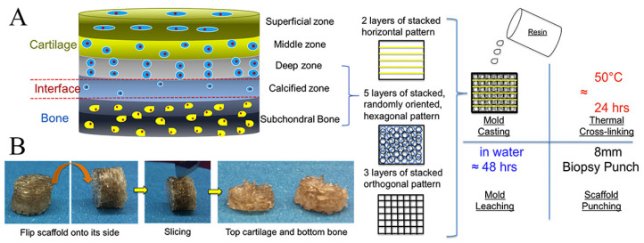
Figure 6.
Schematic illustration of multi-layered scaffold and fabrication designed to emulate the structure of OC tissue by 3D printing technique. Reproduced with permission from Ref. [146]. Copyright 2020, Elsevier. (A) The superficial region contains horizontally aligned cells and fibers and is represented by horizontal fibers in the FDM mold; the intermediate region containing randomly oriented cells and fibers is represented by a randomly oriented hexagonal pore structure; and the deep region contains vertically aligned cells and fibers represented by orthogonal fibers in the FDM mold. (B) Photographic images depicting the resultant scaffold and the sliced bi-layered structure.

