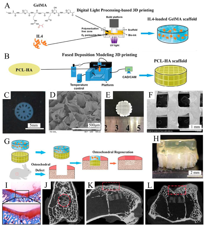
Figure 7.
Fabrication and characterization of the upper GelMA layer and the lower PCL-HA layer. Reproduced with permission from Ref. [143]. Copyright 2020, Elsevier. (A) Schematic of IL-4-loaded GelMA scaffold prepared by the DLP 3D printing system. (B) Schematic of the PCL-HA scaffold prepared by the FDM 3D printing system. (C) Macroscopic image of the GelMA scaffold. (D) SEM images of the GelMA scaffold. (E) Macroscopic image of PCL-HA scaffolds. (F) SEM image of PCL-HA scaffolds (×50). (G) Schematic of fabricating an IL-4-loaded bi-layered scaffold for rabbit osteochondral regeneration. (H) The overall view of the IL-4-loaded bi-layer scaffold. (I) Safranine O staining of the repaired cartilage after 16 weeks post-operation. (J–L) Micro-CT images on the x axis (J), y axis (K), and z axis (L) of the articular joint (n = 3 joints) after operation for 16 weeks.

