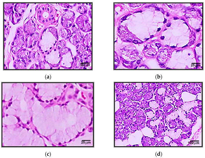
Figure 4.
(a) Microscopic appearance of the submandibular gland with the presence of serous and mixed acini (200xH&E). (b) Coexistence of serous and mucous portions in the same acini (crescent) in the sub-mandibular gland (400xH&E). (c) Microscopic appearance of an area with mucous acini in the submandibular gland (400xH&E). (d) Microscopic appearance of the submandibular gland in the elderly with an increasing amount of adipose tissue (100xH&E).

