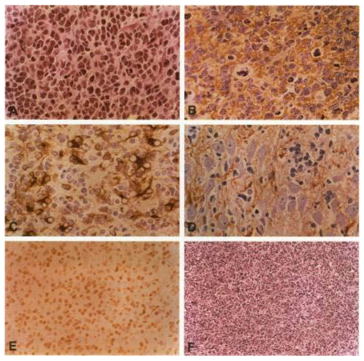
Figure 1.
The rat tumor model of PNETs is histologically indistinguishable from human medulloblastoma. Histology of tumors induced in CNS transplants by retroviral gene expression of SV40 large T antigen. (A) HE-stain with typical histology, incl. neuroblastic Homer-Wright rosettes as a sign of an early stage neuronal differentiation. (B) SYNAPTOPHYSIN expression: immunohistochemical staining with polyclonal antibodies shows a strong synaptophysin expression, indicating neuronal differentiation, (C) GFAP expression: astrocytic differentiation of a tumor cell cluster. (D) Massive infiltration of tumor cells into the hippocampus of the adjacent host brain. Immunohistochemical staining for synaptophysin. (E) Immunohistochemical detection of SV40 large T antigen in the tumor tissue. The monoclonal antibody Pab108 to large T is used for the reaction. Note the characteristic nuclear staining pattern and the absence of immunoreactivity in capillary endothelial cells. (F) Secondary transplant obtained after intracerebral injection of a tumor-derived cell line. The typical morphology is completely preserved. (reproduced/adapted from Eibl et al., 1994 [14]).

