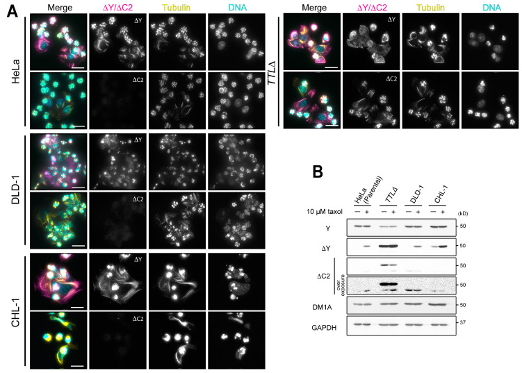
Figure 2.
Taxol treatment increases the levels of ΔY-α-tubulin but not ΔC2-α-tubulin in cells. (A) Detection of ∆Y- and ∆C2-α-tubulin in taxol-treated HeLa, DLD-1, CHL-1, and TTL∆ cells by immunofluorescence (A) and immunoblotting in whole cell lysates (B). In merged images in (A), ∆Y- or ∆C2-α-tubulin is shown in magenta, total α-tubulin (DM1A staining) in yellow, and DNA (DAPI staining) in cyan. Scale bars in (A), 25 µm.

