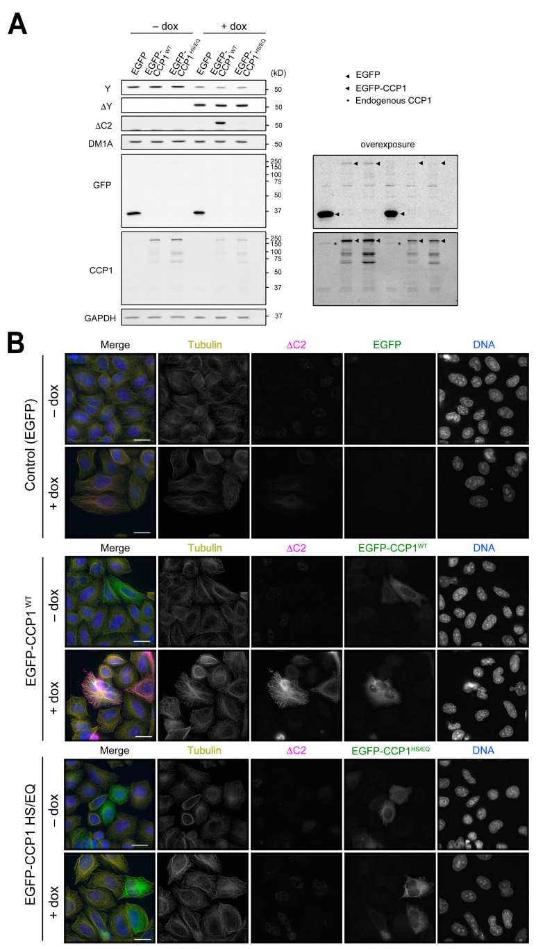
Figure 5.
CCP1 uses ∆Y-α-tubulin as a substrate to generate ∆C2-α-tubulin in cells. (A,B) Detection of ∆C2-α-tubulin in HeLa cells expressing VASH1-SVBP in a doxycycline dependent manner by immunoblotting of cell lysates (A) and immunofluorescence (B). In merged images in (B), total α-tubulin (DM1A staining) is shown in yellow, ∆C2-α-tubulin in magenta, EGFP in green, and DNA (DAPI staining) in blue. Scale bars in (B), 20 µm.

